Epigastric longitudinal scanning
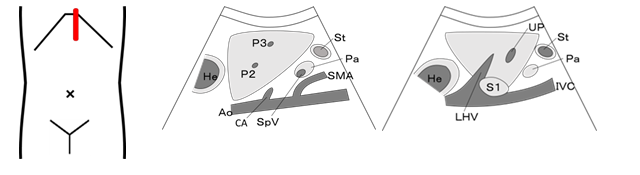
Liver: Caudate lobe(S1), Left lobe (S2), (S3), Quadrate lobe(S4), Portal vein: (P2), (P3), (Umbilical portion; UP), Left hepatic vein; LHV, Aorta; Ao, Inferior vena cava; IVC, Celiac artery; CA, Superior mesenteric artery; SMA, Superior mesenteric vein; SMV, Splenic vein; SpV, Pancreas; Pa, Stomach; St, Heart; He
■Scanning method
Place the probe vertically on the area slightly to the left of the subxiphoid region, and visualize the abdominal aorta. Visualize the lateral segment of the hepatic left lobe on the ventral part of the abdominal aorta, and continue scanning using the fan-like scanning approach until the liver becomes invisible on the scanned image. Similarly, continue scanning the quadrate lobe until the inferior vena cava becomes invisible on the scanned image.
■Precautions
Tilt the probe sufficiently because the upper lateral segment tends to be a blind spot.
If continuity with the previous scan is admitted, perform the scan by moving the probe on the same track as previously done. Be cautions not to rotate the probe 180 degrees.
|
Left subcostal scanning
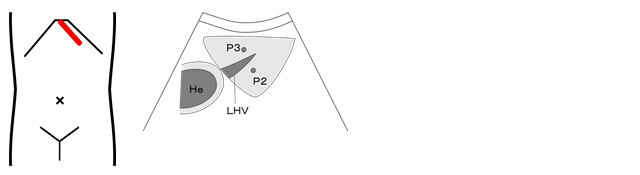
Liver: Left lobe (S2), (S3), Portal vein: (P2), (P3), Left hepatic vein; LHV
■Scanning method
Place the probe along the left sub-costal margin, and continue scanning using the fan-like scanning approach until the hepatic left lobe becomes invisible on the scanned image.
|
Epigastric transverse scanning
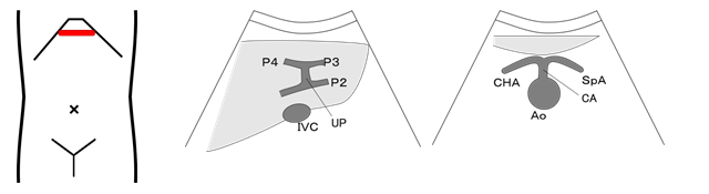
Liver: Caudate lobe(S1), Left lobe(S2), (S3), Quadrate lobe(S4), Portal vein: (P2),(P3), (P4), (Umbilical portion; UP), Left Hepatic vein; LHV, Aorta; Ao, Inferior vena cava; IVC, Celiac artery; CA, Superior mesenteric artery; SMA, Superior mesenteric vein; SMV, Common hepatic artery; CHA, Splenic artery; SpA, Splenic vein; SpV, Pancreas; Pa
■Scanning method
Place the probe horizontally in the epigastric region, and continue scanning using the fan-like scanning approach until the hepatic left lobe becomes invisible on the scanned image.
|
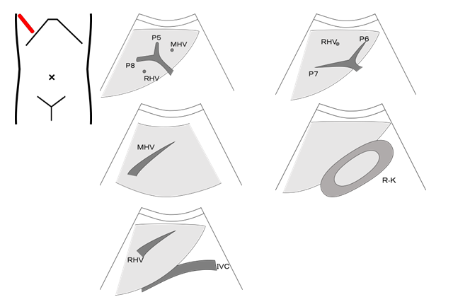
Right subcostal scanning
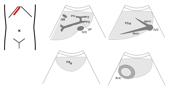
Liver: Right lobe(S5),(S6), (S7), (S8), Right portal vein: (P5), (P6), (P7), (P8), Right hepatic vein; RHV, Middle hepatic vein; MHV, Inferior vena cava; IVC, Gall bladder; GB
Right kidney; R-K, Right adrenal gland
■Scanning method
Place the probe along the right sub-costal margin, and continue scanning using the fan-like scanning approach and the parallel scanning approach until the hepatic right lobe becomes invisible on the scanned image. In addition, move the probe gradually towards the right side along the right subcostal margin, and repeat the same scanning procedure.
■Precautions
Tilt the probe sufficiently because the right margin tends to be a blind spot. Perform the scan with the left lateral decubitus position if visualization is difficult. The cranial part of the right hepatic lobe, which is also called the "dome," tends to be a blind spot.
|
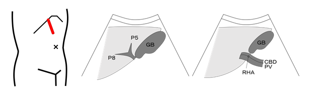
Right hypochondrium longitudinal scanning

Gall bladder; GB, Common bile duct; CBD, Liver: Quadrate lobe(S4), Right lobe(S5), (S8), Right portal vein: (P5), (P8), Right kidney; R-K
■Scanning method
Place the probe vertically in the right hypochondrium, and visualize a long-axis cross-sectional view of the gall bladder.
Continue scanning using the fan-like scanning approach and the parallel scanning approach until the gall bladder becomes invisible on the scanned image.
■Precautions
Perform the scan using the left lateral decubitus position if visualization is difficult.
|
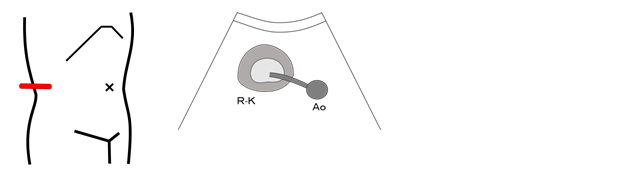
Right intercostal scanning
Liver: Right lobe(S5),(S6), (S7), (S8), Right portal vein: (P5), (P6), (P7), (P8), Right hepatic vein; RHV, Middle hepatic vein; MHV, Inferior vena cava; IVC, Gall bladder; GB
Right kidney; R-K, Morrison's pouch, Right adrenal gland
■Scanning method
Place the probe on the right intercostal space, and continue scanning each intercostal region using the fan-like scanning approach until the right hepatic lobe becomes invisible on the scanned image.
Place the probe as far dorsally as possible during visualization of the long-axis cross-section of the right kidney, and scan the site by tilting the ultrasound beam anteriorly.
■Precautions
Observe the right subdiaphragmatic region with the patient controlling their breathing.
The adrenal glands are usually surrounded by hyperechoic fatty tissues and connective tissues and are often difficult to visualize.
|
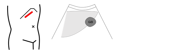
Right hypochondrium diagonal scanning
Liver, Common bile duct; CBD, Gall bladder; GB, Right hepatic artery; RHA, Portal vein; PV, Pancreas head, Duodenum
■Scanning method
Place the probe obliquely in the right hypochondrium, and observe the courses of the portal vein and the common bile duct located in the ventral part of the portal vein.
■Precautions
Left lateral decubitus position.
|
Right hypochondrium transverse scanning
Liver, Common bile duct; CBD, Gall bladder; GB, Right hepatic artery; RHA, Portal vein; PV, Pancreas head, Duodenum
■Scanning method
Place the probe vertically in the right hypochondrium, and visualize a long-axis cross-sectional view of the gall bladder.
Continue scanning using the fan-like scanning approach and the parallel scanning approach until the gall bladder becomes invisible on the scanned image.
|
Right longitudinal scanning
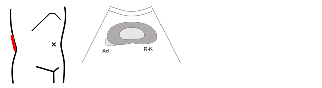
Right kidney; R-K, Medulla, Cortex, Renal pelvis, Ureter, Renal sinus, Adrenal grand; Ad
■Scanning method
Visualize the long axis view of the right kidney in its right dorsal aspect, and continue scanning using the fan-like scanning approach until the kidney becomes invisible on the scanned image.
■Precautions
The adrenal glands are usually surrounded by hyperechoic fatty tissues and are often difficult to visualize.
|
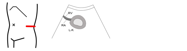
Right transverse scanning
Right kidney; R-K, Ureter, Renal Artery; RA, Renal Vein; RV, Aorta; Ao
■Scanning method
Continue to scan the site from the upper pole to the lower pole with the probe rotated 90 degrees counterclockwise as viewed from the long axis of the right kidney, using the fan-like scanning approach and the parallel scanning approach until the kidney becomes invisible on the scanned image.
|
Left intercostal scanning
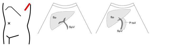
Spleen; Sp, Splenic artery; SpA, Splenic vein; SpV, Left kidney; L-K, Pancreas tail; P-tail, Adrenal gland; Ad
■Scanning method
Place the probe on the left intercostal space, and then move it dorsally.
Continue scanning each intercostal area using the fan-like scanning approach until the spleen becomes invisible on the scanned image.
■Precautions
Visualize the spleen by moving the probe ventrally while scanning.
Observe the left subdiaphragmatic region with the patient controlling his/her breathing.
The tail of the pancreas can easily be identified and visualized after the splenic vein is visualized, using the spleen as an acoustic window.
|
Left longitudinal scanning
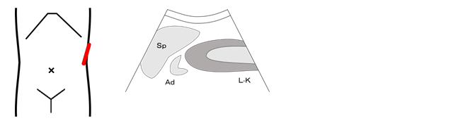
Spleen; Sp, Left kidney; L-K, Adrenal gland; Ad
■Scanning method
Scan the site in the same manner as that used for the right adrenal gland, while visualizing the long-axis cross-section of the left kidney.
|
Left longitudinal scanning
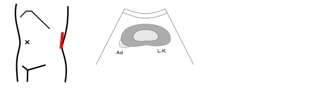
Left kidney; L-K, Medulla, Cortex, Renal pelvis, Ureter, Renal sinus, Adrenal grand; Ad
■Scanning method
Visualize the long axis view of the left kidney in its left dorsal aspect, and continue scanning using the fan-like scanning approach until the kidney becomes invisible on the scanned image.
■Precautions
Right lateral decubitus position.
|
Left transverse scanning
Left kidney; L-K, Medulla, Cortex, Renal pelvis, Ureter, Renal sinus, Adrenal grand; Ad
■Scanning method
Scan the site in the same manner as that used for the right kidney with the probe rotated 90 degrees counterclockwise as viewed from the long axis of the left kidney.
|
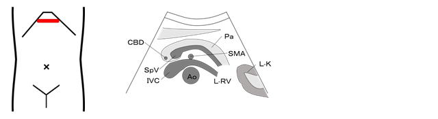
Epigastric longitudinal scanning
Pancreas; Pa, Aorta; Ao, Inferior vena cava; IVC, Superior mesenteric artery; SMA, Superior mesenteric vein; SMV, Celiac artery; CA, Splenic artery; SpA, Splenic vein; SpV, Stomach; St, Common bile duct; CBD, Liver: left lobe
■Scanning method
Place the probe in the median sagittal plane in the subxiphoid region, and visualize the abdominal aorta and the superior mesenteric artery. Continue scanning the region from the pancreatic head to the tail, centering on the body of the pancreas at the ventral part of the bifurcation of the superior mesenteric artery, using the fan-like scanning approach and the parallel scanning approach until the pancreas becomes invisible on the scanned image.
■Precautions
Perform the scan after the patient has had a drink of water, or in the sitting position if visualization is difficult.
|
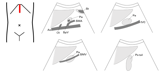
Epigastric transverse scanning
Pancreas; Pa, Aorta; Ao, Inferior vena cava; IVC, Superior mesenteric artery; SMA, Superior mesenteric vein; SMV, Celiac artery; CA, Splenic vein; SpV, Stomach; St, Common bile duct; CBD, Left renal vein; L-RV, Stomach; St, Common bile duct; CBD
Liver, left lobe: Left adrenal
■Scanning method
Rotate the probe approximately 90 degrees counterclockwise as viewed from the position where the pancreatic body is visualized on the longitudinal scan, and obtain a long axis view of the pancreas. Continue scanning using the fan-like scanning approach and the parallel scanning approach until the pancreas becomes invisible on the scanned image.
■Precautions
Visualize the pancreas using the liver as an acoustic window.
If visualization is difficult because of gastrointestinal gases or for some other reason, use a seated position or an approach that involves filling the stomach.
In a thin person, visualization becomes difficult if the pancreas is strongly compressed by the probe.
Left oblique scanning is required to obtain a long axis view of the pancreas.
|
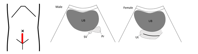
Transverse scanning
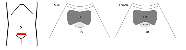
Male
Urinary bladder; UB(top, body, fundus, neck), Prostate; Pr, Seminal vesicle, Ureter; Ure
Female
Urinary bladder; UB(top, body, fundus, neck), Uterus; Ut, Vagina; VA, Ovary, Ureter; Ure
■Scanning method
Place the probe horizontally on the upper margin of the pubis, and continue scanning from top to bottom, using the fan-like scanning approach and the parallel scanning approach until the urinary bladder becomes invisible on the scanned image.
For observation of the urinary bladder neck, place the probe on the upper margin of the pubis, and tilt the ultrasound beam towards direction of the feet.
■Precautions
The bladder lumen tends to be affected by artifacts such as multiple reflections and side lobes.
|
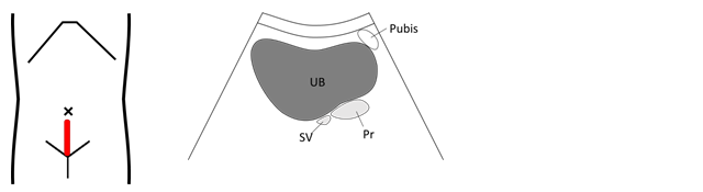
Longitudinal scanning
Male
Urinary bladder; UB(top, body, fundus, neck), Prostate; Pr, Seminal vesicle; SV, Ureter; Ure
Female
Urinary bladder; UB(top, body, fundus, neck), Uterus; Ut, Vagina; Va, Ovary, Ureter; Ure
■Scanning method
Place the probe vertically on the upper margin of the pubis, and continue scanning using the fan-like scanning approach and the parallel scanning approach until the urinary bladder becomes invisible on the scanned image.
|
Right diagonal scanning
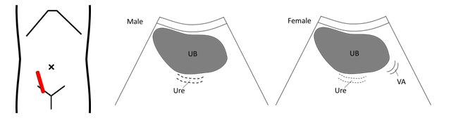
Male
Ureter; Ure, Ureter pelvic junction, Urinary bladder; UB(top, body, fundus, neck), Prostate; Pr, Seminal vesicle; SV
Female
Ureter; Ure, Ureter pelvic junction, Urinary bladder; UB(top, body, fundus, neck), Uterus; Ut, Vagina; VA, Ovary
■Scanning method
Observe the right Vesicoureteral junction with the probe rotated approximately 45 degrees counterclockwise as viewed from the median long axis of the urinary bladder.
|
Left diagonal scanning
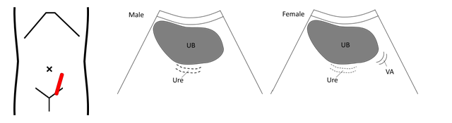
Male
Urinary bladder; UB(top, body, fundus, neck), Ureter; Ure, Ureter pelvic junction, Prostate; Pr, Seminal vesicle; SV
Female
Urinary bladder; UB(top, body, fundus, neck), Ureter; Ure, Ureter pelvic junction, Uterus; Ut, Ovary
■Scanning method
Observe the left Vesicoureteral junction with the probe rotated approximately 45 degrees clockwise as viewed from the median long axis of the urinary bladder.
|
Transverse scanning
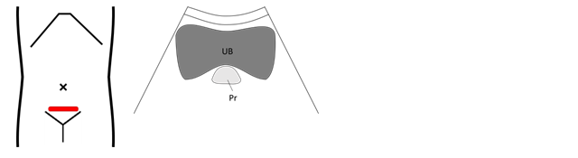
Male
Urinary bladder; UB(neck, fundus), Prostate(innergland, outergland); Pr, Seminal vesicle; SV, Ureter; Ure
■Scanning method
Place the probe horizontally on the upper margin of the pubis, and move the ultrasound beam in a continuous, caudo-cranial direction, using the fan-like scanning approach until the prostate gland becomes invisible on the scanned image.
■Precautions
Excessive filling of the urinary bladder may compress the prostate gland dorsally, causing shape changes or poor visualization.
|
Longitudinal scanning
Male
Urinary bladder; UB(neck, bottom), Prostate(innergland, outergland); Pr, Seminal vesicle; SV, Ureter; Ure
■Scanning method
Place the probe vertically so that it touches the upper margin of the pubis, and continue scanning using the fan-like scanning approach and the parallel scanning approach until the prostate gland becomes invisible on the scanned image.
|
Preparative regimen: Full urinary bladder technique should be employed. When the test is after discharging urine, give the patient 500~600ml of water until he/she has a desire to urinate.
|
Longitudinal scanning
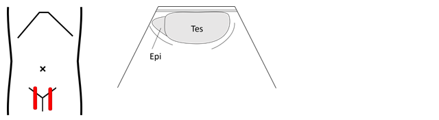
Male
Testis; Tes, Epididymis; Epi, Seminal vesicle; SV, Upper body of semina, Vesicle, Spermatic cord vein, Scrotal skin
■Scanning method
Place the probe along the long axis of the testis, and continue scanning using the parallel scanning approach until the testis becomes invisible on the scanned image.
■Precautions
This test becomes easier with the penis elevated anteriorly.
|
Transverse scanning
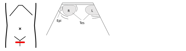
Male
Testis; Tes, Epididymis; Epi, Seminal vesicle; SV, Upper body of semina, Vesicle, Spermatic cord vein, Scrotal skin
■Scanning method
Continue scanning from the upper pole to the lower pole of the testis until the testis becomes invisible on the scanned image, with the probe rotated 90 degrees counterclockwise as viewed from the long-axis cross-section of the testis.
|
Preparative regimen: High-frequency wave should be used for the probe. The test becomes easier with the probe having a wide bore and a light-weighed small water bag directly attached.
|
◇This is the secondary use with a partial revision of the text translated under the responsibility of the Ministry of Economy, Trade and Industry.